 中文
中文 别名:Sciellin, SCEL应用:WB,IHC,FCM
反应种属:Human
规格:50μl/100μl
| Description |
|---|
| The protein encoded by this gene is a precursor to the cornified envelope of terminally differentiated keratinocytes. This protein localizes to the periphery of cells and may function in the assembly or regulation of proteins in the cornified envelope. Transcript variants encoding different isoforms exist. A transcript variant utilizing an alternative polyA signal has been described in the literature, but its full-length nature has not been determined. |
| Specification | |
|---|---|
| Aliases | Sciellin, SCEL |
| Entrez GeneID | 8796 |
| Swissprot | O95171 |
| WB Predicted band size | 77.6kDa |
| Host/Isotype | Rabbit IgG |
| Storage | Store at 4°C short term. Aliquot and store at -20°C long term. Avoid freeze/thaw cycles. |
| Species Reactivity | Human |
| Immunogen | This SCEL antibody is generated from rabbits immunized with a KLH conjugated synthetic peptide between 260-289 amino acids from the Central region of human SCEL. |
| Formulation | Purified polyclonal antibody supplied in PBS with 0.05% sodium azide. This antibody is purified through a protein A column, followed by peptide affinity purification. |
| Application | |
|---|---|
| WB | 1/1000 |
| IHC | 1/500 |
| FCM | 1/25 |
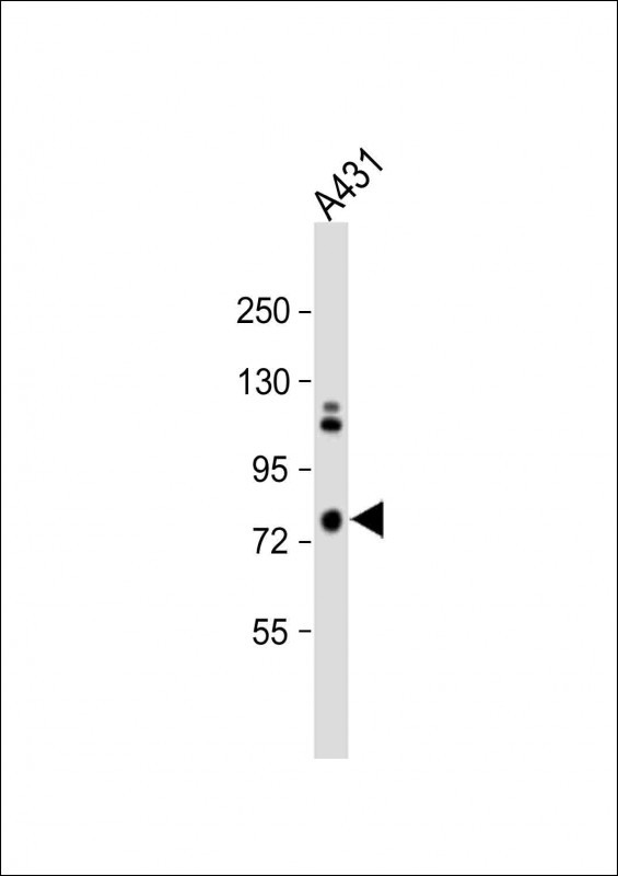 |
Anti-SCEL Antibody (Center) at 1:1000 dilution + A431 whole cell lysate
Lysates/proteins at 20 µg per lane. Secondary Predicted band size : 78 kDa Blocking/Dilution buffer: 5% NFDM/TBST. |
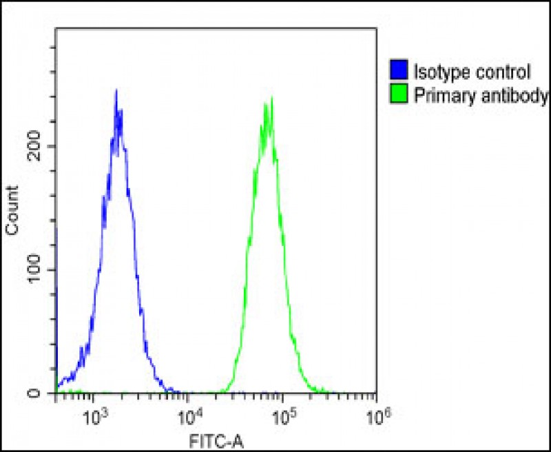 |
Overlay histogram showing A431 cells stained with P34599(green line). The cells were fixed with 2% paraformaldehyde and then permeabilized with 90% methanol for 10 min. The cells were then icubated in 2% bovine serum albumin to block non-specific protein-protein interactions followed by the antibody (1:25 dilution) for 60 min at 37ºC. The secondary antibody used was Goat-Anti-Rabbit IgG, DyLight® 488 Conjugated Highly Cross-Adsorbed at 1/200 dilution for 40 min at Room temperature. Isotype control antibody (blue line) was rabbit IgG1 (1μg/1×10^6 cells) used under the same conditions. Acquisition of >10, 000 events was performed. |
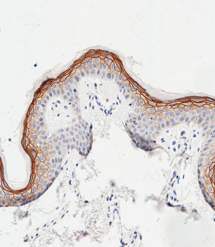 |
Immunohistochemical analysis of paraffin-embedded human skin tissue using P34599 performed on the Leica® BOND RXm. Samples were incubated with primary antibody(1/500) for 1 hours at room temperature. A undiluted biotinylated CRF Anti-Polyvalent HRP Polymer antibody was used as the secondary antibody. |
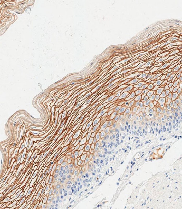 |
Immunohistochemical analysis of paraffin-embedded human esophagus tissue using P34599 performed on the Leica® BOND RXm. Samples were incubated with primary antibody(1/500) for 1 hours at room temperature. A undiluted biotinylated CRF Anti-Polyvalent HRP Polymer antibody was used as the secondary antibody. |
本公司的所有产品仅用于科学研究或者工业应用等非医疗目的,不可用于人类或动物的临床诊断或治疗,非药用,非食用。
暂无评论
本公司的所有产品仅用于科学研究或者工业应用等非医疗目的,不可用于人类或动物的临床诊断或治疗,非药用,非食用。


发表回复