 中文
中文 别名:Serum deprivation-response protein, Cavin-2, PS-p68, Phosphatidylserine-binding protein, SDPR {ECO:0000312|EMBL:AAD177951}应用:WB,IHC,FCM
反应种属:Human
规格:50μl/100μl
| Description |
|---|
| This gene encodes a calcium-independent phospholipid-binding protein whose expression increases in serum-starved cells. This protein is a substrate for protein kinase C (PKC) phosphorylation and recruits polymerase I and transcript release factor (PTRF) to caveolae. Removal of this protein causes caveolae loss and its over-expression results in caveolae deformation and membrane tubulation. |
| Specification | |
|---|---|
| Aliases | Serum deprivation-response protein, Cavin-2, PS-p68, Phosphatidylserine-binding protein, SDPR {ECO:0000312|EMBL:AAD177951} |
| Entrez GeneID | 8436 |
| Swissprot | O95810 |
| WB Predicted band size | 47.2kDa |
| Host/Isotype | Rabbit IgG |
| Storage | Store at 4°C short term. Aliquot and store at -20°C long term. Avoid freeze/thaw cycles. |
| Species Reactivity | Human |
| Immunogen | This SDR antibody is generated from rabbits immunized with a KLH conjugated synthetic peptide between 109-135 amino acids from the Central region of human SDR. |
| Formulation | Purified polyclonal antibody supplied in PBS with 0.05% sodium azide. This antibody is prepared by Saturated Ammonium Sulfate (SAS) precipitation followed by dialysis against PBS. |
| Application | |
|---|---|
| WB | 1/1000 |
| IHC | 1/100-1/500 |
| FCM | 1/10-1/50 |
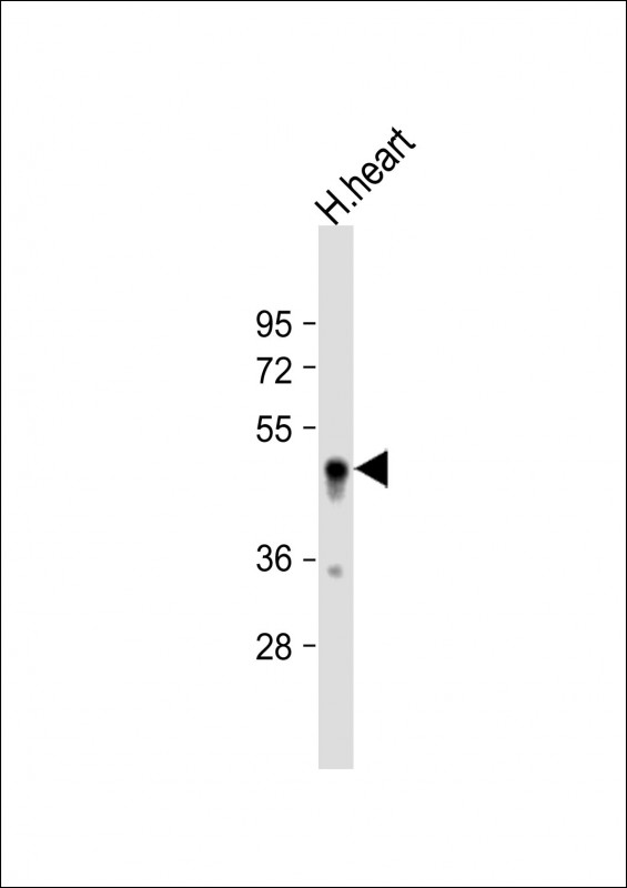 |
Anti-SDR Antibody (Center) at 1:1000 dilution + human heart lysate
Lysates/proteins at 20 µg per lane. Secondary Predicted band size : 47 kDa Blocking/Dilution buffer: 5% NFDM/TBST. |
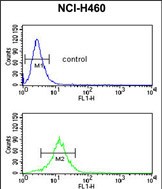 |
SDR Antibody (Center) (Cat. #P33455) flow cytometric analysis of NCI-H460 cells (bottom histogram) compared to a negative control cell (top histogram).FITC-conjugated goat-anti-rabbit secondary antibodies were used for the analysis. |
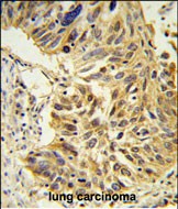 |
SDR Antibody (Center) (Cat. #P33455) IHC analysis in formalin fixed and paraffin embedded lung carcinoma followed by peroxidase conjugation of the secondary antibody and DAB staining. This data demonstrates the use of the SDR Antibody (Center) for immunohistochemistry. Clinical relevance has not been evaluated. |
本公司的所有产品仅用于科学研究或者工业应用等非医疗目的,不可用于人类或动物的临床诊断或治疗,非药用,非食用。
暂无评论
本公司的所有产品仅用于科学研究或者工业应用等非医疗目的,不可用于人类或动物的临床诊断或治疗,非药用,非食用。


发表回复