 中文
中文 别名:E3 ubiquitin-protein ligase SMURF2, hSMURF2, 632-, SMAD ubiquitination regulatory factor 2, SMAD-specific E3 ubiquitin-protein ligase 2, SMURF2应用:WB,IHC,ICC
反应种属:Human, Mouse, Rat
规格:50μl/100μl
| Description |
|---|
| SMURF2 is an E3 ubiquitin-protein ligase which accepts ubiquitin from an E2 ubiquitin-conjugating enzyme in the form of a thioester and then directly transfers the ubiquitin to targeted substrates. This protein interacts with SMAD1, SMAD2 and SMAD7 in order to trigger their ubiquitination and proteasome-dependent degradation. It enhances the inhibitory activity of SMAD7 and reduces the transcriptional activity of SMAD2. Coexpression of SMURF2 with SMAD1 results in considerable decrease in steady-state level of SMAD1 protein and a smaller decrease of SMAD2 level. |
| Specification | |
|---|---|
| Aliases | E3 ubiquitin-protein ligase SMURF2, hSMURF2, 632-, SMAD ubiquitination regulatory factor 2, SMAD-specific E3 ubiquitin-protein ligase 2, SMURF2 |
| Entrez GeneID | 64750 |
| Swissprot | Q9HAU4 |
| WB Predicted band size | 86.2kDa |
| Host/Isotype | Rabbit IgG |
| Storage | Store at 4°C short term. Aliquot and store at -20°C long term. Avoid freeze/thaw cycles. |
| Species Reactivity | Human, Mouse, Rat |
| Immunogen | This SMURF2 antibody is generated from rabbits immunized with a KLH conjugated synthetic peptide between 702-731 amino acids from the C-terminal region of human SMURF2. |
| Formulation | Purified polyclonal antibody supplied in PBS with 0.05% sodium azide. This antibody is prepared by Saturated Ammonium Sulfate (SAS) precipitation followed by dialysis against PBS. |
| Application | |
|---|---|
| WB | 1/1000-1/2000 |
| IHC | 1/100-1/500 |
| ICC | 1/10-1/50 |
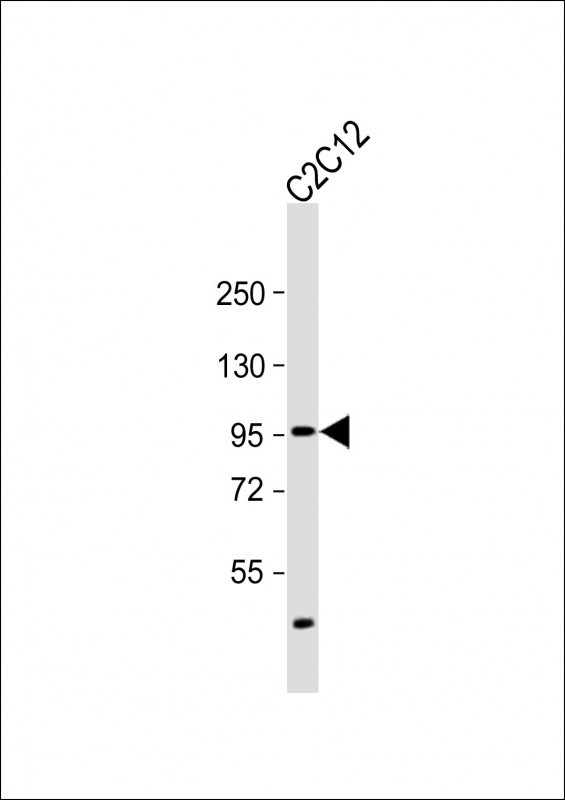 |
Anti-SMURF2 Antibody (C-term) at 1:2000 dilution + C2C12 whole cell lysate
Lysates/proteins at 20 µg per lane. Secondary Predicted band size : 86 kDa Blocking/Dilution buffer: 5% NFDM/TBST. |
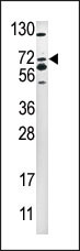 |
Western blot analysis of anti-SMURF2 Pab (Cat. #P33924) in 293 cell line lysate (35ug/lane). SMURF2(arrow) was detected using the purified Pab |
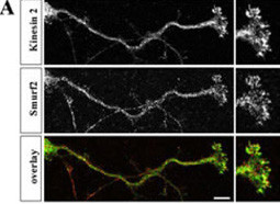 |
Hippocampal neurons were fixed at stage 3, stained with anti-Smurf2 (red) and anti-Kinesin-2 (green) antibodies, and analyzed by confocal microscopy. The panels show single confocal planes. (J. Biol. Chem. 2007 Nov 30;282(48):35259-35268) |
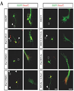 |
Hippocampal neurons were transfected 2 h after plating with expression vectors for EGFP, EGFP-tagged Par3-4N/2, Par3-PDZ2, Par3-PDZ3, Smurf2-HECT (HECT), Smurf2-HECT-C716A (HECT CA), and shRNA directed against mPar3 (Par3 RNAi), or vectors for the anti-Par3 shRNA and human Myc-Par3 (RNAi + h Par3) (green). Transfected cells were analyzed at 3 d.i.v. by staining with an anti-Smurf2 antibody (red). Axons are marked by arrowheads. The marked growth cones are shown at a higher magnification. Scale bars, 40 and 10 ?. (J. Biol. Chem. 2007 Nov 30;282(48):35259-35268) |
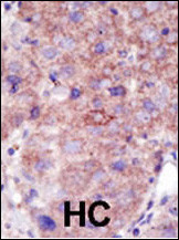 |
Formalin-fixed and paraffin-embedded human cancer tissue reacted with the primary antibody, which was peroxidase-conjugated to the secondary antibody, followed by DAB staining. This data demonstrates the use of this antibody for immunohistochemistry; clinical relevance has not been evaluated. BC = breast carcinoma; HC = hepatocarcinoma. |
本公司的所有产品仅用于科学研究或者工业应用等非医疗目的,不可用于人类或动物的临床诊断或治疗,非药用,非食用。
暂无评论
本公司的所有产品仅用于科学研究或者工业应用等非医疗目的,不可用于人类或动物的临床诊断或治疗,非药用,非食用。


发表回复