 中文
中文 别名:Small nuclear ribonucleoprotein Sm D3, Sm-D3, snRNP core protein D3, SNRPD3应用:WB,IHC
反应种属:Human, Mouse
规格:50μl/100μl
| Description |
|---|
| The protein encoded by this gene belongs to the small nuclear ribonucleoprotein core protein family. It is required for pre-mRNA splicing and small nuclear ribonucleoprotein biogenesis. |
| Specification | |
|---|---|
| Aliases | Small nuclear ribonucleoprotein Sm D3, Sm-D3, snRNP core protein D3, SNRPD3 |
| Entrez GeneID | 6634 |
| Swissprot | P62318 |
| WB Predicted band size | 13.9kDa |
| Host/Isotype | Rabbit IgG |
| Storage | Store at 4°C short term. Aliquot and store at -20°C long term. Avoid freeze/thaw cycles. |
| Species Reactivity | Human, Mouse |
| Immunogen | This SNRPD3 antibody is generated from rabbits immunized with a KLH conjugated synthetic peptide between 9-38 amino acids from the N-terminal region of human SNRPD3. |
| Formulation | Purified polyclonal antibody supplied in PBS with 0.05% sodium azide. This antibody is purified through a protein A column, followed by peptide affinity purification. |
| Application | |
|---|---|
| WB | 1/2000-1/4000 |
| IHC | 1/500 |
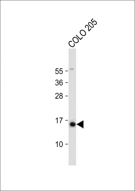 |
Anti-SNRPD3 Antibody (N-term) at 1:2000 dilution + COLO 205 whole cell lysate
Lysates/proteins at 20 µg per lane. Secondary Predicted band size : 14 kDa Blocking/Dilution buffer: 5% NFDM/TBST. |
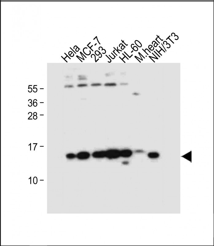 |
All lanes : Anti-SNRPD3 Antibody (N-term) at 1:4000 dilution Lane 1: Hela whole cell lysate Lane 2: MCF-7 whole cell lysate Lane 3: 293 whole cell lysate Lane 4: Jurkat whole cell lysate Lane 5: HL-60 whole cell lysate Lane 6: Mouse heart tissue lysate Lane 7: NIH/3T3 whole cell lysate Lysates/proteins at 20 µg per lane. Secondary Predicted band size : 14 kDa Blocking/Dilution buffer: 5% NFDM/TBST. |
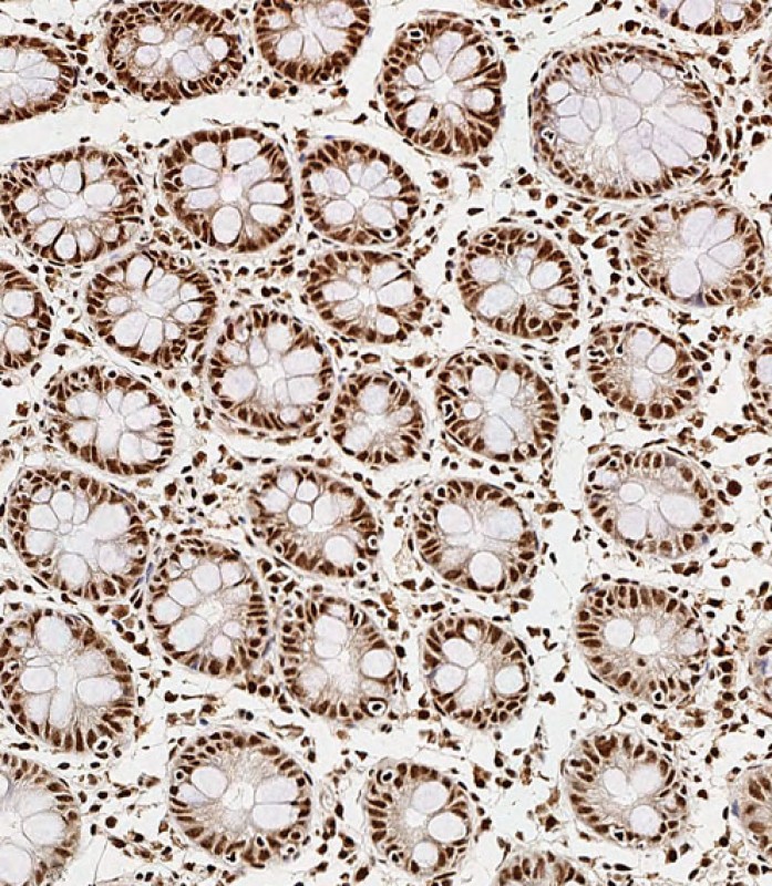 |
Immunohistochemical analysis of paraffin-embedded Human colon tissue using P34687 performed on the Leica® BOND RXm. Tissue was fixed with formaldehyde at room temperature, antigen retrieval was by heat mediation with a EDTA buffer (pH9. 0). Samples were incubated with primary antibody(1:500) for 1 hours at room temperature. A undiluted biotinylated CRF Anti-Polyvalent HRP Polymer antibody was used as the secondary antibody. |
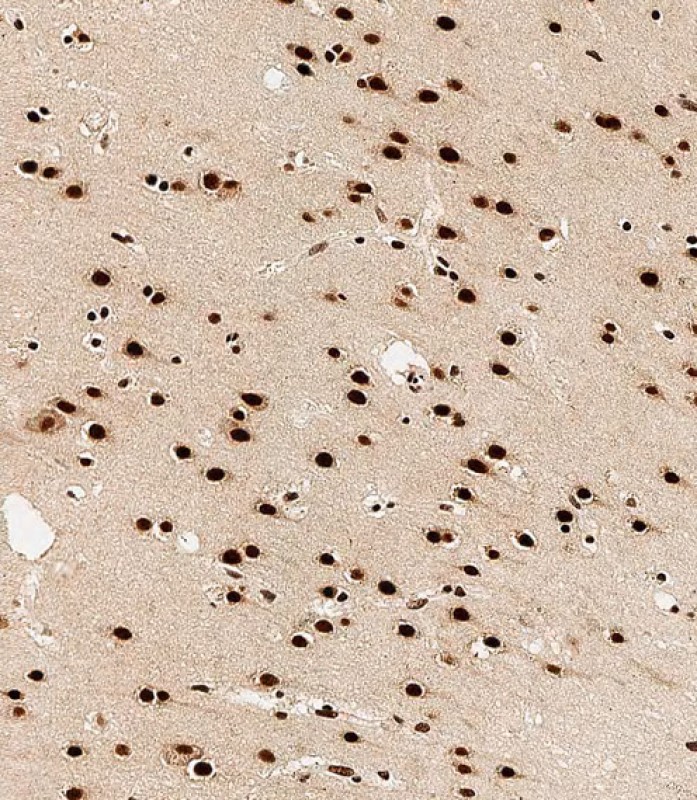 |
Immunohistochemical analysis of paraffin-embedded Human brain tissue using P34687 performed on the Leica® BOND RXm. Tissue was fixed with formaldehyde at room temperature, antigen retrieval was by heat mediation with a EDTA buffer (pH9. 0). Samples were incubated with primary antibody(1:500) for 1 hours at room temperature. A undiluted biotinylated CRF Anti-Polyvalent HRP Polymer antibody was used as the secondary antibody. |
本公司的所有产品仅用于科学研究或者工业应用等非医疗目的,不可用于人类或动物的临床诊断或治疗,非药用,非食用。
暂无评论
本公司的所有产品仅用于科学研究或者工业应用等非医疗目的,不可用于人类或动物的临床诊断或治疗,非药用,非食用。


发表回复