 中文
中文 别名:Protein spinster homolog 2, SPNS2应用:WB,FCM
反应种属:Human, Mouse
规格:50μl/100μl
| Description |
|---|
| Sphingolipid transporter required for migration of myocardial precursors. Transports sphingosine 1-phosphate (S1P), a secreted lipid mediator that plays critical roles in cardiovascular, immunological, and neural development and function. Mediates the export of S1P from cells in the extraembryonic yolk syncytial layer (YSL), thereby regulating myocardial precursor migration. |
| Specification | |
|---|---|
| Aliases | Protein spinster homolog 2, SPNS2 |
| Entrez GeneID | 124976 |
| Swissprot | Q8IVW8 |
| WB Predicted band size | 58.0kDa |
| Host/Isotype | Rabbit IgG |
| Storage | Store at 4°C short term. Aliquot and store at -20°C long term. Avoid freeze/thaw cycles. |
| Species Reactivity | Human, Mouse |
| Immunogen | This SPNS2 antibody is generated from rabbits immunized with a KLH conjugated synthetic peptide between 68-94 amino acids from the N-terminal region of human SPNS2. |
| Formulation | Purified polyclonal antibody supplied in PBS with 0.05% sodium azide. This antibody is purified through a protein A column, followed by peptide affinity purification. |
| Application | |
|---|---|
| WB | 1/1000 |
| FCM | 1/25 |
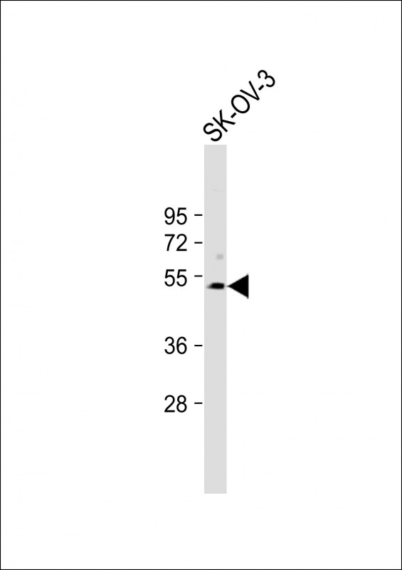 |
Anti-SPNS2 Antibody (N-term) at 1:1000 dilution + SK-OV-3 whole cell lysate
Lysates/proteins at 20 µg per lane. Secondary Predicted band size : 58 kDa Blocking/Dilution buffer: 5% NFDM/TBST. |
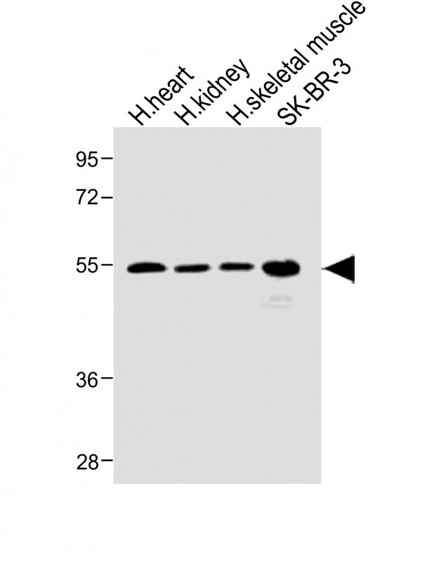 |
All lanes : Anti-SPNS2 Antibody (N-term) at 1:1000 dilution Lane 1: Human heart lysate Lane 2: Human kidney lysate Lane 3: Human skeletal muscle lysate Lane 4: SK-BR-3 whole cell lysate Lysates/proteins at 20 µg per lane. Secondary Predicted band size : 58 kDa Blocking/Dilution buffer: 5% NFDM/TBST. |
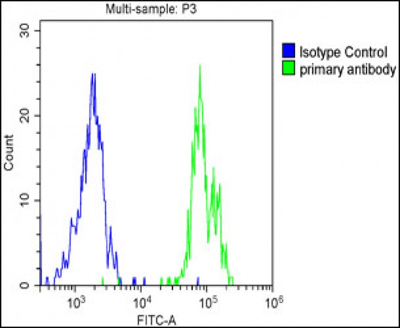 |
Overlay histogram showing SK-OV-3 cells stained with P34593(green line). The cells were fixed with 2% paraformaldehyde and then permeabilized with 90% methanol for 10 min. The cells were then icubated in 2% bovine serum albumin to block non-specific protein-protein interactions followed by the antibody (1:25 dilution) for 60 min at 37ºC. The secondary antibody used was Goat-Anti-Rabbit IgG, DyLight® 488 Conjugated Highly Cross-Adsorbed at 1/200 dilution for 40 min at Room temperature. Isotype control antibody (blue line) was rabbit IgG1 (1μg/1×10^6 cells) used under the same conditions. Acquisition of >10, 000 events was performed. |
本公司的所有产品仅用于科学研究或者工业应用等非医疗目的,不可用于人类或动物的临床诊断或治疗,非药用,非食用。
暂无评论
本公司的所有产品仅用于科学研究或者工业应用等非医疗目的,不可用于人类或动物的临床诊断或治疗,非药用,非食用。


发表回复