 中文
中文 别名:Sulfotransferase 1A1, ST1A1, Aryl sulfotransferase 1, HAST1/HAST2, Phenol sulfotransferase 1, Phenol-sulfating phenol sulfotransferase 1, P-PST 1, ST1A3, Thermostable phenol sulfotransferase, Ts-PST, SULT1A1, STP, STP1应用:WB,FCM
反应种属:Human
规格:50μl/100μl
| Description |
|---|
| Sulfotransferase that utilizes 3′-phospho-5′-adenylyl sulfate (PAPS) as sulfonate donor to catalyze the sulfate conjugation of catecholamines, phenolic drugs and neurotransmitters. Has also estrogen sulfotransferase activity. responsible for the sulfonation and activation of minoxidil. Is Mediates the metabolic activation of carcinogenic N- hydroxyarylamines to DNA binding products and could so participate as modulating factor of cancer risk. |
| Specification | |
|---|---|
| Aliases | Sulfotransferase 1A1, ST1A1, Aryl sulfotransferase 1, HAST1/HAST2, Phenol sulfotransferase 1, Phenol-sulfating phenol sulfotransferase 1, P-PST 1, ST1A3, Thermostable phenol sulfotransferase, Ts-PST, SULT1A1, STP, STP1 |
| Entrez GeneID | 6817 |
| Swissprot | P50225 |
| WB Predicted band size | 34.2kDa |
| Host/Isotype | Rabbit IgG |
| Storage | Store at 4°C short term. Aliquot and store at -20°C long term. Avoid freeze/thaw cycles. |
| Species Reactivity | Human |
| Immunogen | This SULT1A1 antibody is generated from a rabbit immunized with a KLH conjugated synthetic peptide between 246-279 amino acids of human SULT1A1. |
| Formulation | Purified polyclonal antibody supplied in PBS with 0.05% sodium azide. This antibody is purified through a protein A column, followed by peptide affinity purification. |
| Application | |
|---|---|
| WB | 1/1000 |
| FCM | 1/25 |
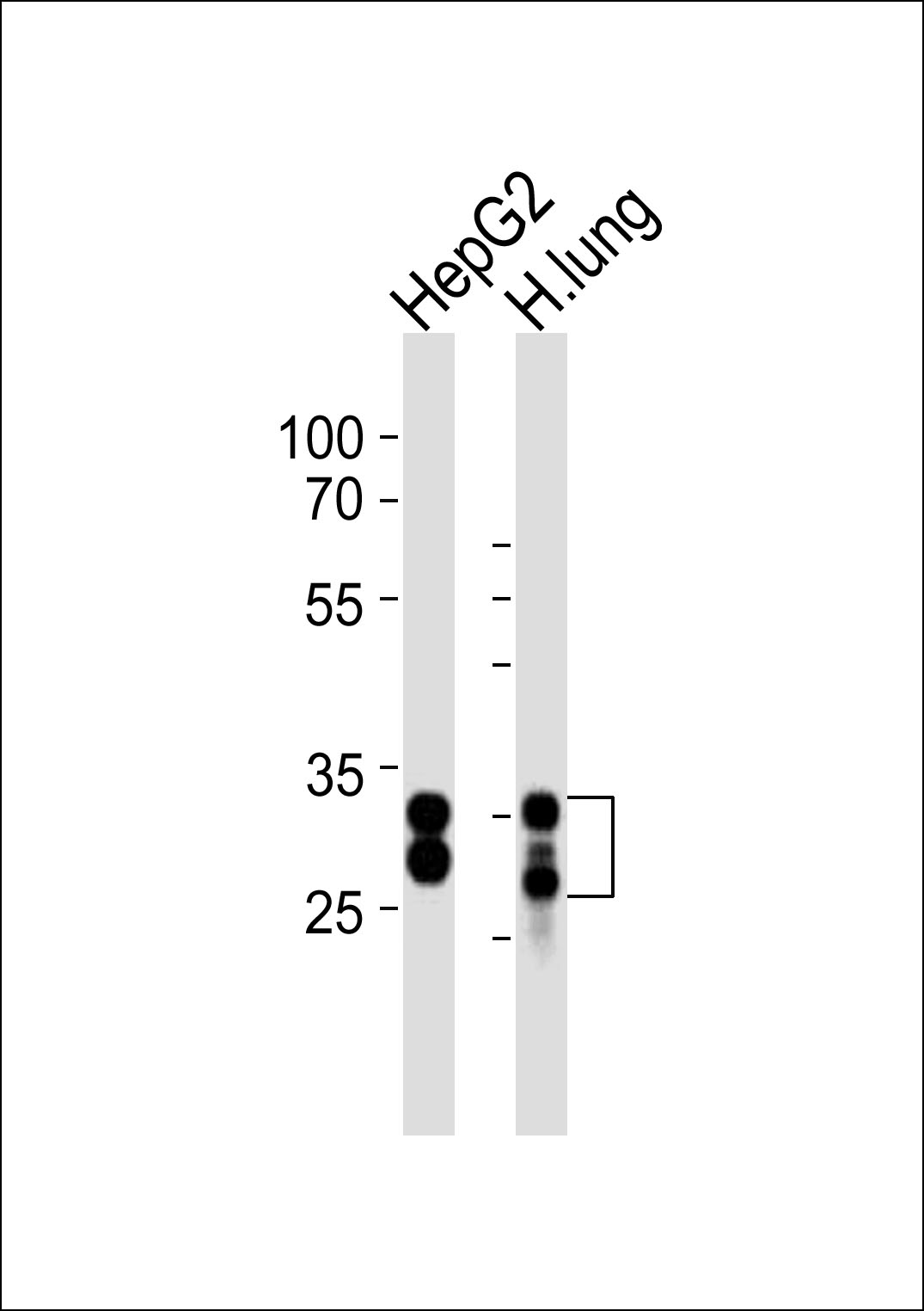 |
Western blot analysis of lysates from HepG2 cell line and human lung tissue (from left to right), using SULT1A1 Antibody (C-term)(Cat. #P32920). P32920 was diluted at 1:1000 at each lane. A goat anti-rabbit IgG H&L(HRP) at 1:10000 dilution was used as the secondary antibody. Lysates at 20ug per lane. |
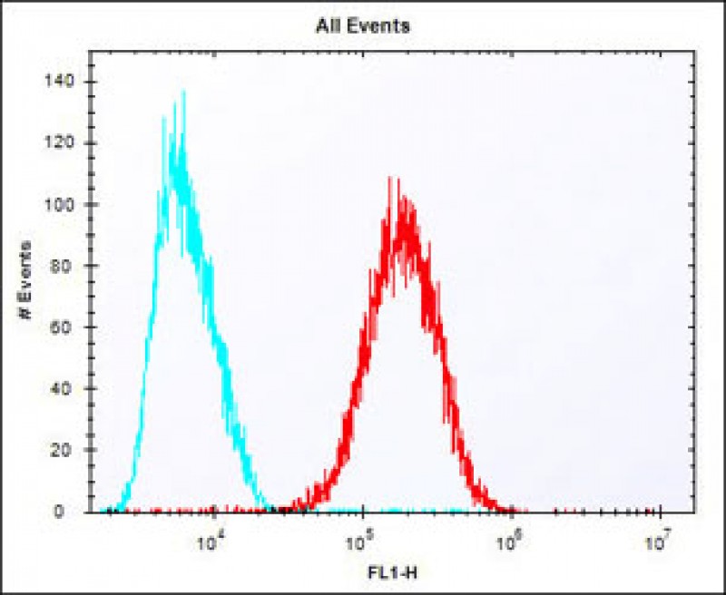 |
Overlay histogram showing HepG2 cells stained with P32920 (red line). The cells were fixed with 2% paraformaldehyde (10 min) and then permeabilized with 90% methanol for 10 min. The cells were then icubated in 2% bovine serum albumin to block non-specific protein-protein interactions followed by the antibody (P32920, 1:25 dilution) for 60 min at 37ºC. The secondary antibody used was Alexa Fluor® 488 goat anti-rabbit lgG (H+L) (1583138) at 1/400 dilution for 40 min at 37ºC. Isotype control antibody (blue line) was rabbit IgG1 (1μg/1×10^6 cells) used under the same conditions. Acquisition of >10, 000 events was performed. |
本公司的所有产品仅用于科学研究或者工业应用等非医疗目的,不可用于人类或动物的临床诊断或治疗,非药用,非食用。
暂无评论
本公司的所有产品仅用于科学研究或者工业应用等非医疗目的,不可用于人类或动物的临床诊断或治疗,非药用,非食用。


发表回复