 中文
中文 别名:E3 ubiquitin-protein ligase synoviolin, 632-, Synovial apoptosis inhibitor 1, SYVN1, HRD1, KIAA1810应用:WB,IHC,ICC
反应种属:Human, Mouse
规格:50μl/100μl
| Description |
|---|
| HRD1 is a ubiquitin ligase whose expression is induced by the unfolded protein response (UPR) following endoplasmic reticulum stress. Expression of HRD1 protects cells from apoptosis by inducing degradation of abnormally processed proteins that accumulate in the endoplasmic reticulum. HRD1 is expressed in many tissues, strongly expressed in brain, pancreas, liver, kidney and skeletal muscle. Amano T, et al. reported that Synoviolin/Hrd1 (expressed in rheumatoid synovium) is a novel causative factor for arthropathy by triggering synovial cell outgrowth through its antiapoptotic effects. HRD1 contains one ring-type zinc finger. |
| Specification | |
|---|---|
| Aliases | E3 ubiquitin-protein ligase synoviolin, 632-, Synovial apoptosis inhibitor 1, SYVN1, HRD1, KIAA1810 |
| Entrez GeneID | 84447 |
| Swissprot | Q86TM6 |
| WB Predicted band size | 67.7kDa |
| Host/Isotype | Rabbit IgG |
| Storage | Store at 4°C short term. Aliquot and store at -20°C long term. Avoid freeze/thaw cycles. |
| Species Reactivity | Human, Mouse |
| Immunogen | This SYVN1 (HRD1) antibody is generated from rabbits immunized with a KLH conjugated synthetic peptide between 586-617 amino acids from the C-terminal region of human SYVN1 (HRD1). |
| Formulation | Purified polyclonal antibody supplied in PBS with 0.05% sodium azide. This antibody is purified through a protein A column, followed by peptide affinity purification. |
| Application | |
|---|---|
| WB | 1/1000-1/2000 |
| IHC | 1/100-1/500 |
| ICC | 1/200 |
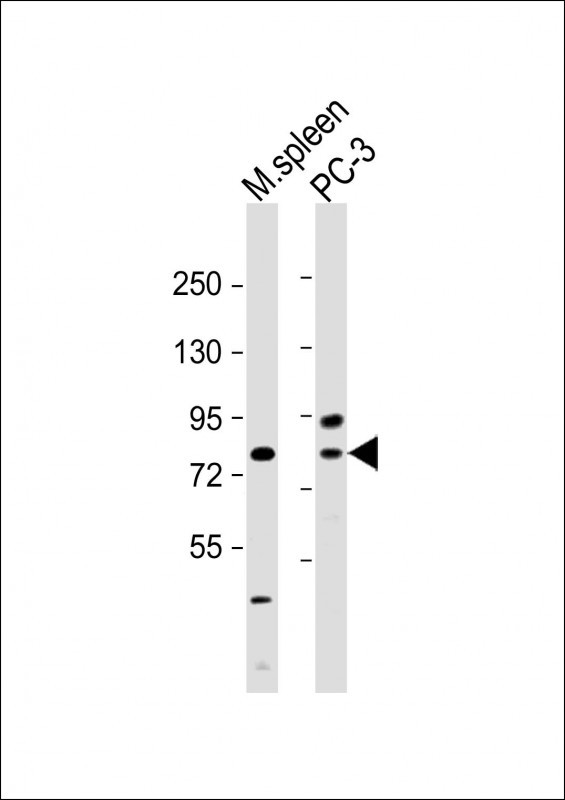 |
All lanes : Anti-HRD1 Antibody (A601) at 1:2000 dilution Lane 1: mouse spleen lysate Lane 2: PC-3 whole cell lysate Lysates/proteins at 20 µg per lane. Secondary Predicted band size : 68 kDa Blocking/Dilution buffer: 5% NFDM/TBST. |
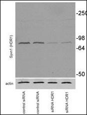 |
Mouse Neuroblastoma Neuro2A (N2A) was transiently transfected, collected at 72h after transfection. Primary antibodies against syvn1 (Abgent # P34352, 1:1000) and anti-rabbit secondary POD-conjugated antibodies from Pierce Biotechnology, Inc (Rockford, IL, 1:2000)(Provided by Dr. Susana Granell & Institution University of Arkansas). |
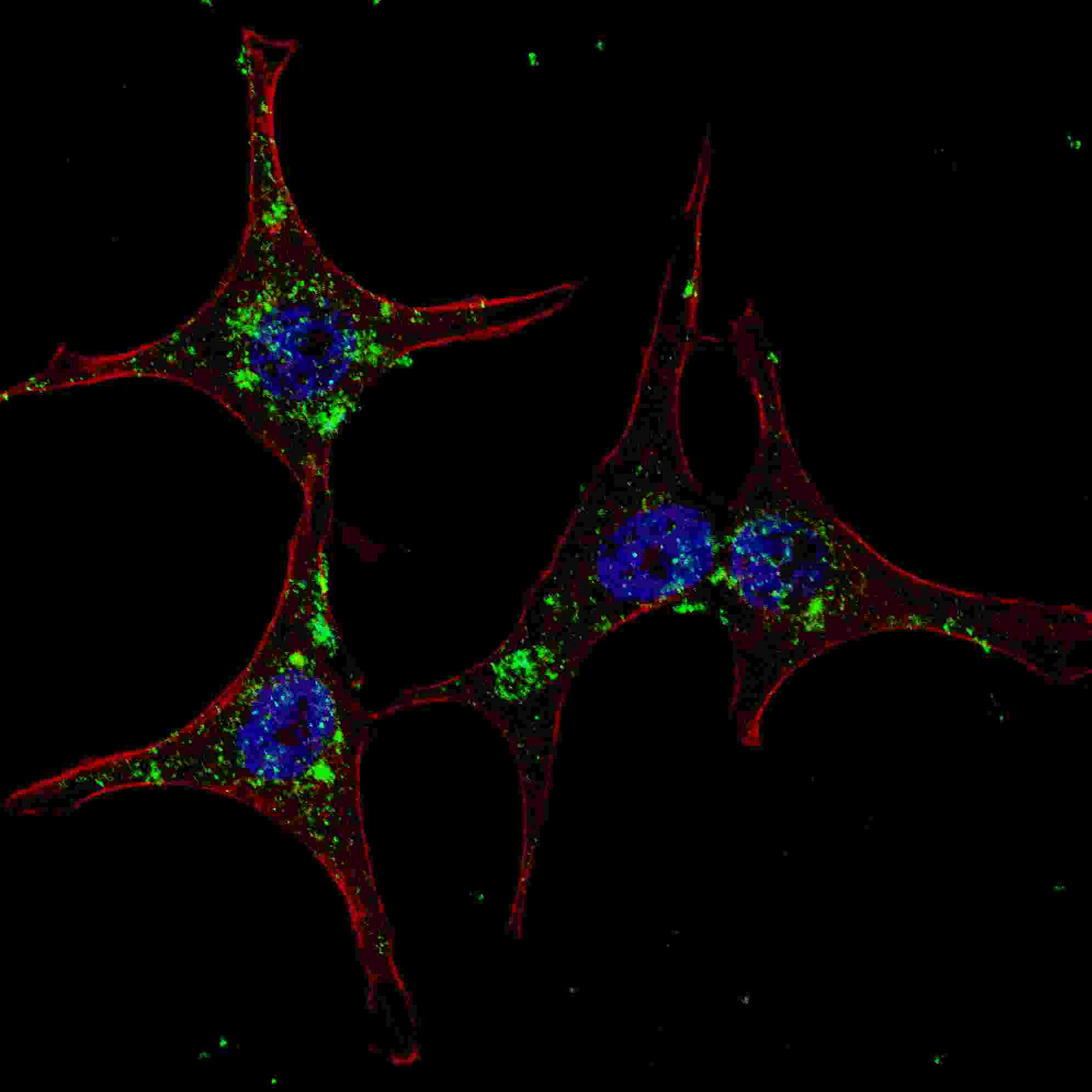 |
Fluorescent confocal image of HeLa cells stained with SYVN1 (HRD1) (C-term) antibody. HeLa cells were fixed with 4% PFA (20 min), permeabilized with Triton X-100 (0.2%, 30 min). Cells were then incubated with P34352 SYVN1 (HRD1) (C-term) primary antibody (1:200, 2 h at room temperature). For secondary antibody, Alexa Fluor® 488 conjugated donkey anti-rabbit antibody (green) was used (1:1000, 1h). Nuclei were counterstained with Hoechst 33342 (blue) (10 μg/ml, 5 min). |
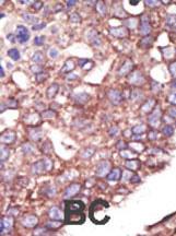 |
Formalin-fixed and paraffin-embedded human cancer tissue reacted with the primary antibody, which was peroxidase-conjugated to the secondary antibody, followed by DAB staining. This data demonstrates the use of this antibody for immunohistochemistry; clinical relevance has not been evaluated. BC = breast carcinoma; HC = hepatocarcinoma. |
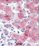 |
Formalin-fixed and paraffin-embedded human Liver tissue reacted with SYVN1 (HRD1) Antibody (C-term)(Cat.#P34352), which was peroxidase-conjugated to the secondary antibody, followed by AEC staining. This data demonstrates the use of this antibody for immunohistochemistry; clinical relevance has not been evaluated. |
本公司的所有产品仅用于科学研究或者工业应用等非医疗目的,不可用于人类或动物的临床诊断或治疗,非药用,非食用。
暂无评论
本公司的所有产品仅用于科学研究或者工业应用等非医疗目的,不可用于人类或动物的临床诊断或治疗,非药用,非食用。


发表回复