 中文
中文 别名:Wilms tumor protein, WT33, WT1应用:WB,IHC,ICC,FCM
反应种属:Human, Mouse, Rat
规格:50μl/100μl
| Description |
|---|
| This gene encodes a transcription factor that contains four zinc-finger motifs at the C-terminus and a proline/glutamine-rich DNA-binding domain at the N-terminus. It has an essential role in the normal development of the urogenital system, and it is mutated in a small subset of patients with Wilm’s tumors. This gene exhibits complex tissue-specific and polymorphic imprinting pattern, with biallelic, and monoallelic expression from the maternal and paternal alleles in different tissues. Multiple transcript variants have been described. In several variants, there is evidence for the use of a non-AUG (CUG) translation initiation site upstream of and in-frame with the first AUG. Authors of PMID:7926762 also provide evidence that WT1 mRNA undergoes RNA editing in human and rat, and that this process is tissue-restricted and developmentally regulated. [provided by RefSeq]. |
| Specification | |
|---|---|
| Aliases | Wilms tumor protein, WT33, WT1 |
| Entrez GeneID | 7490 |
| Swissprot | P19544 |
| WB Predicted band size | 49.2kDa |
| Host/Isotype | Rabbit IgG |
| Storage | Store at 4°C short term. Aliquot and store at -20°C long term. Avoid freeze/thaw cycles. |
| Species Reactivity | Human, Mouse, Rat |
| Immunogen | This WT1 antibody is generated from rabbits immunized with a KLH conjugated synthetic peptide between 346-375 amino acids from the Central region of human WT1. |
| Formulation | Purified polyclonal antibody supplied in PBS with 0.05% sodium azide. This antibody is purified through a protein A column, followed by peptide affinity purification. |
| Application | |
|---|---|
| WB | 1/1000 |
| IHC | 1/100-1/500 |
| ICC | 1/10-1/50 |
| FCM | 1/10-1/50 |
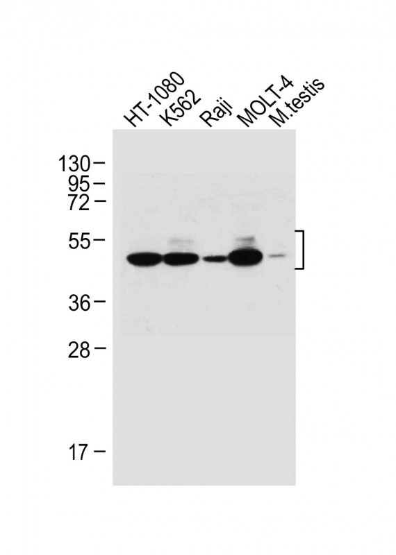 |
All lanes : Anti-WT1 Antibody (Center E361) at 1:1000 dilution Lane 1: HT-1080 whole cell lysate Lane 2: K562 whole cell lysate Lane 3: Raji whole cell lysate Lane 4: MOLT-4 whole cell lysate Lane 5: Mouse testis lysate Lysates/proteins at 20 µg per lane. Secondary Predicted band size : 49 kDa Blocking/Dilution buffer: 5% NFDM/TBST. |
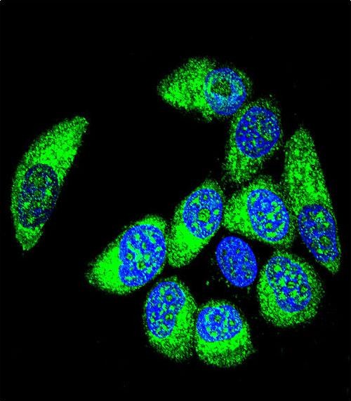 |
Confocal immunofluorescent analysis of WT1 Antibody (Center E361)(Cat. #P34639) with MCF-7 cell followed by Alexa Fluor® 488-conjugated goat anti-rabbit lgG (green). DAPI was used to stain the cell nuclear (blue). |
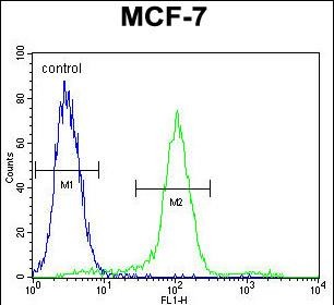 |
WT1 Antibody (Center E361) (Cat. #P34639) flow cytometric analysis of MCF-7 cells (right histogram) compared to a negative control cell (left histogram).FITC-conjugated goat-anti-rabbit secondary antibodies were used for the analysis. |
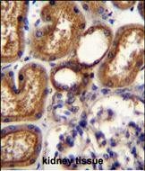 |
WT1 Antibody (Center E361) (Cat. #P34639)immunohistochemistry analysis in formalin fixed and paraffin embedded human kidney tissue followed by peroxidase conjugation of the secondary antibody and DAB staining.This data demonstrates the use of WT1 Antibody (Center E361) for immunohistochemistry. Clinical relevance has not been evaluated. |
本公司的所有产品仅用于科学研究或者工业应用等非医疗目的,不可用于人类或动物的临床诊断或治疗,非药用,非食用。
暂无评论
本公司的所有产品仅用于科学研究或者工业应用等非医疗目的,不可用于人类或动物的临床诊断或治疗,非药用,非食用。


发表回复